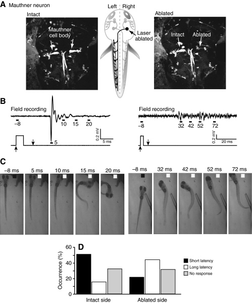
Fig. 7.
Unilateral Mauthner ablation impairs fast escape and associated electric field potentials. (A) Confocal images of dye-filled Mauthner cell bodies were obtained before (left) and immediately after (right) laser ablation of the right Mauthner cell performed at 4 d.p.f. Schematic drawing (center) shows approximate Mauthner cell location (arrow). (B) Electric field potentials (top) were recorded in response to stimuli delivered to the intact (left) or ablated (right) sides at 5 d.p.f. The lower trace corresponds to the Picospritzer pulse. (C) Selected frames show behavioral responses evoked by stimuli delivered to the intact (left) or ablated (right) sides. Behavioral responses correspond with the field potentials shown in B. Representative results are shown in B and C. (D) Percentage of short- and long-latency scape responses to stimuli delivered to the intact and ablated sides. Results were obtained from nine animals in which 81 and 94 stimuli were applied to the intact and ablated sides, respectively. Gray bars indicate the percentage of trials that failed to elicit escape behavior. Included in this category are trials in which the animal righted itself after being knocked over by the stimulus jet.

