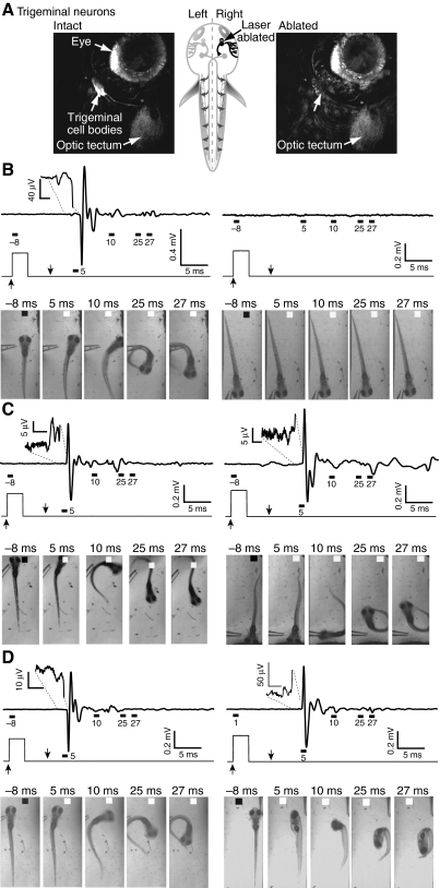
Fig. 8.
Unilateral ablation of trigeminal neurons impairs fast and slow escapes and associated electric field potentials. (A) Confocal images of GFP-expressing trigeminal sensory neurons were obtained before (left) and immediately after (right) laser ablation of neurons in the right trigeminal ganglion performed at 3 d.p.f. Unlabelled arrow in right panel points to area previously occupied by trigeminal cell bodies. Schematic drawing (center) shows approximate location of a trigeminal neuron (arrow). (B–D) Results were obtained 1 (B), 3 (C), or 7 (D) days post-ablation (d.p.a.) from four (B,C) or two animals (D). Top: electric field potentials were recorded in response to stimuli delivered to the intact (left) or ablated (right) sides. Insets show the initial, neurogenic component of the signal on an expanded scale. Middle: the Picospritzer pulse. Bottom: selected frames show behavioral responses evoked by stimuli delivered to the intact (left) or ablated (right) sides. In C (3 d.p.a.), the animal turns toward the stimulus rather than away in response to a stimulus delivered to the ablated side. Field potentials and the corresponding behavioral responses were recorded in the same trial. Results are representative of data obtained from four animals at 1 and 3 d.p.a. and from two animals at 7 d.p.a. Day 1: 23 stimuli on intact side, 31 stimuli on ablated side; day 3: 18 stimuli on intact side, 21 stimuli on ablated side; day 7, 15 stimuli on intact side, 19 stimuli on ablated side.

