Figure 4.
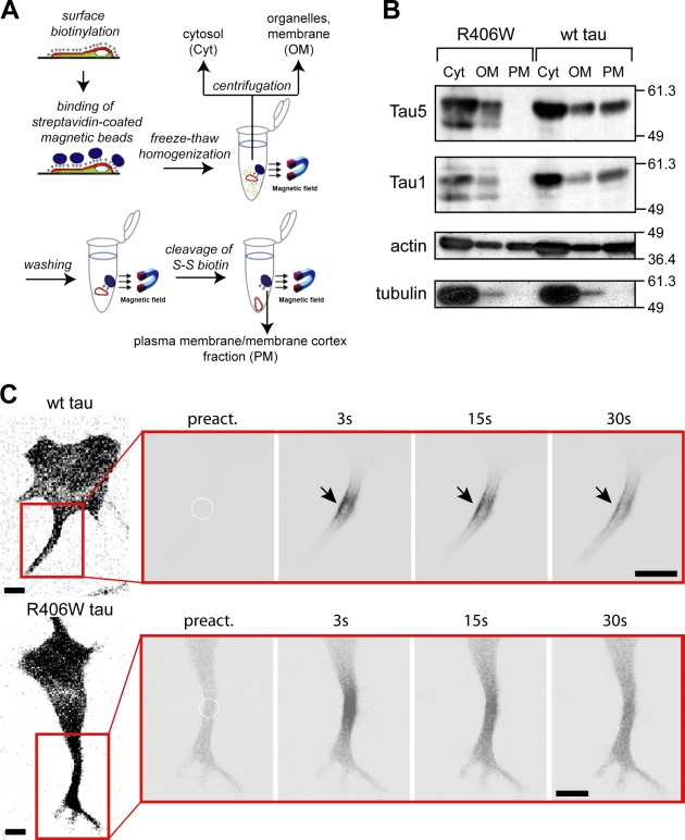
R406W tau is deficient in binding to the neural plasma membrane. (A) Schematic representation of the plasma membrane fractionation assay to analyze tau’s interaction with the neural membrane cortex. (B) Immunoblot showing the distribution of FLAG-tagged tau, actin, and tubulin in the cytosolic (Cyt), organelles/membrane (OM), and plasma membrane/membrane cortex (PM) fractions. Note the complete absence of R406W tau mutant in the PM fraction, whereas a major amount of wt tau is PM-associated. Numbers to the sides of the gel blots indicate molecular mass standards in kilodaltons. (C) Time-lapse microscopic images of processes from PC12 cells expressing PAGFP-tagged tau constructs after photoactivation. A close-up of the processes (red box) is shown and the position of photoactivation is indicated by a circle. Note the enrichment of wt tau close to the plasma membrane (arrows), whereas R406W tau shows a uniform distribution. Experiments were performed with the 352 tau isoform. Bar, 10 µm.

