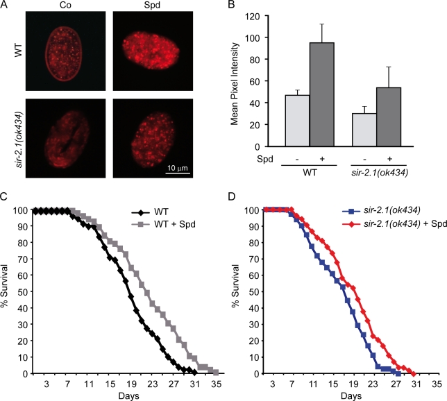
Figure 3.
The life-extending and autophagy-inducing effects of spermidine in C. elegans are not mediated by Sir2. (A) Fluorescence microscopy of C. elegans transgenic embryos expressing a full-length plgg-1DsRed::LGG-1 fusion protein indicative of autophagic activity. Two representative pictures of wild-type (WT) and sir-2.1 embryos untreated (Co, control) or treated with 0.2-mM spermidine (Spd) supplementation of food are shown. (B) Quantification of autophagic activity through the measurement of DsRed::LGG-1 pixel intensity from images of WT animals shown in A. Data represent means ± SEM (n = 3) with ≥25 images processed for each trial. (C) Survival of WT C. elegans during aging with and without (control) supplementation of food (UV-killed E. coli) with 0.2-mM spermidine (n = 110; P < 0.005). (D) Survival of sir-2.1 C. elegans (ok434 phenotype) during aging with and without (control) supplementation of food (UV-killed E. coli) with 0.2-mM spermidine (n = 110; P < 0.01). P-values were calculated using the log-rank test as described in Materials and methods.

