Figure 8.
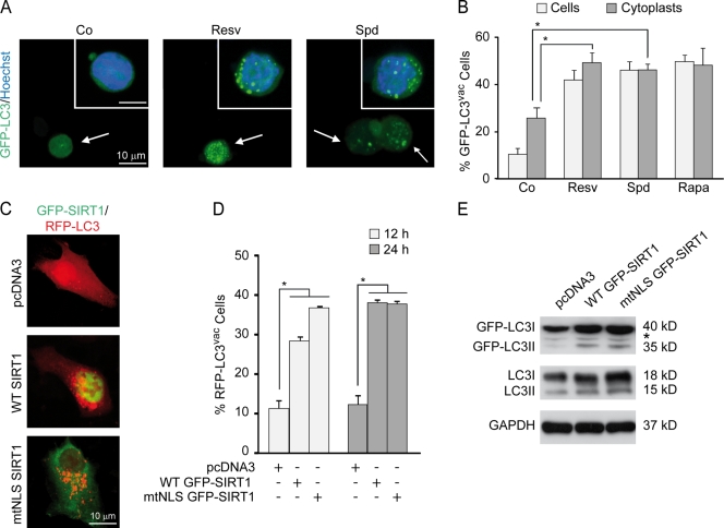
Autophagy can be efficiently regulated by cytoplasmic (de)acetylation reactions. (A and B) HCT 116 cells were transfected with a GFP-LC3–encoding plasmid, cultured in complete medium for 24 h, and enucleated to obtain cytoplasts. The cytoplasts were treated with either vehicle (Co, control), 100-µM resveratrol (Resv), or 100-µM spermidine (Spd) for 4 h. 1-µM rapamycin (Rapa) was used as a positive control. (A) Representative images of cytoplasts indicative of autophagic activity. Arrows indicate GFP-LC3–transfected cytoplasts, whereas insets show nonenucleated transfected cells. (B) Quantitative data. Bars show the percentages of cells or cytoplasts showing the accumulation of GFP-LC3 in puncta (GFP-LC3vac). (C–E) HCT 116 cells were cotransfected with a plasmid for the expression of RFP-LC3 together with an empty vector (pcDNA3) or plasmids encoding wild-type (WT) SIRT1 or a SIRT1 variant with a mutation in the nuclear localization signal fused to GFP (WT GFP-SIRT1 and mtNLS GFP-SIRT1, respectively) for 12 or 24 h, fixed, and analyzed by fluorescence microscopy. (C) Representative images indicative of 24-h autophagic activity. (D) Quantitative data. Bars depict the percentages of cells exhibiting the accumulation of RFP-LC3 in puncta (RFP-LC3vac). (B and D) Means ± SEM; n = 3; *, P < 0.05. (E) Representative immunoblots for 24-h endogenous LC3 lipidation. The asterisk stands for a nonspecific band. GAPDH, glyceraldehyde 3-phosphate dehydrogenase.

