Figure 3.
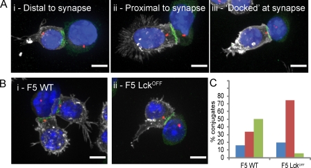
Lck is required for complete polarization and docking of the centrosome to the immunological synapse. (A) Merged immunofluorescence projections of centrosome polarization phenotypes seen in F5 CTL conjugates with the centrosome distal, proximal, or docked at the synapse (i.e., with γ-tubulin contacting the plasma membrane marker CD8), with cells labeled with antibodies against talin (green), γ-tubulin (red), and CD8 (white). Nuclei are stained with Hoechst (blue). (B) Representative images of centrosome polarization observed in WT and Lckoff CTLs. Every image (A and B) is a merge of four channels collected from z stacks. (C) Quantitation of centrosome polarization for F5 WT (n = 112) and F5 Lckoff (n = 103) from data analyzed in 3D showing the percentage of conjugates with the centrosome distal (blue), proximal (red), or docked (green) at the synapse. A two-tailed Student’s t test for loss of centrosome docking in Lckoff samples compared with WT gave a statistical significance of P = 10−4. Centrosome polarization was also quantitated in three independent experiments without 3D reconstruction (Fig. S3). Bars, 5 µm.

