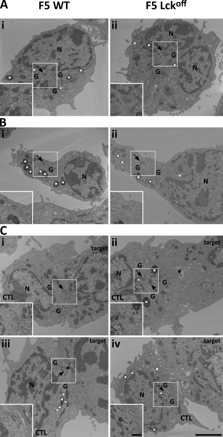
Figure 4.
The centrosome fails to dock at the immunological synapse in Lckoff CTLs. (A–C) EM micrographs from thin (50–100 nm) lead-stained sections of nonmotile (A), motile (B), and target-conjugated (C) F5 WT or Lckoff CTLs loaded with HRP to reveal the endocytic pathway. The Golgi complex (G), lytic granules (white asterisks), secretory cleft (black asterisks), nuclei (N), and centrosome (arrows) are indicated in each image. Insets are magnified images of the centrosome area marked by white boxes. Bars: (main panels) 2 µm; (insets) 0.5 µm.

