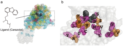
Figure 5. 3D structure of GPCR and a ligand.
(a) 3D structure of GPCR (β2 adrenergic receptor) and a ligand (Carazolol, colored black) derived from PDB, target substructure in GPCR being colored according to the corresponding group of significant substructure pairs. (b) An enlargement of (a), focusing on the binding site with target substructures including DRY (for activation), and CW, LPF, PFF, LVM and SID (for binding), all being in G6.

