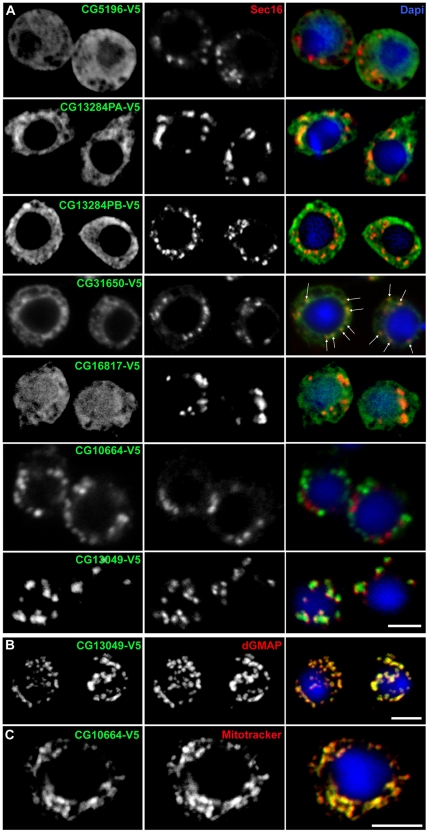
Figure 6. Localisation/overexpression of selected hits.
(A) Immunofluorescence (IF) localisation of six V5-tagged hits in Drosophila S2 cells using an anti-V5 monoclonal antibody (green) and an anti-Sec16 antibody recognizing endogenous Sec16 (red). The merged channels are shown in the third column together with Dapi. Both CG13284 isoforms were expressed and exhibited similar ER localisation. Arrows in CG31650 panel point at puncta of more concentrated staining that partially overlap with Sec16, likely representing tER-Golgi units. (B) IF localisation of CG13049-V5 (green) with respect to the Golgi that is labeled with an anti-dGMAP antibody (red). (C) IF localisation of CG10664-V5 (green) with respect to mitochondria that are stained with mitotracker (red). Note in B and C the complete overlap between the tagged proteins and Golgi membrane and mitochondria, respectively. Representative confocal sections are shown. Scale bars: 5 µm.

