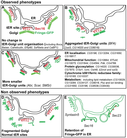
Figure 8. Screen phenotypes.
(A) No change in the organisation of the tER-Golgi units when compared to non depleted cells. The tER sites are depicted with COPII vesicles decorated by red Sec16, and the Golgi apparatus is represented as a green paired Golgi stacks marked by Fringe-GFP [8]. This phenotype was obtained for 2/3 of the depleted genes. (B) Aggregated phenotype: The tER-Golgi units appear aggregated on one side of the cells and seemingly larger. Again, the Golgi apparatus can have retained its wild-type organisation (as in cells treated with BFA, [8]) or be fragmented. This phenotype was observed in 4 out of 49 hits. (C) MG phenotype: the tER sites retain their spatial relationship to the Golgi as in non-depleted cells but they appear smaller. However, whether the two compartments have an altered structure is not assessed. The Golgi apparatus can be a smaller paired stack, or a single stack (as in Abi and Scar depletion; [8]) or fragmented into vesicles and tubules (as in SMSr depletion; [9]). This phenotype is observed in 41 out 49 hits. The most prominent functional groups of proteins, whose depletion leads to this phenotype, are listed. (D) The Golgi is heavily fragmented, retains or not its spatial relationship with the tER sites that are not affected. This phenotype was not observed. (E) Fringe-GFP is retained in the ER and the tER sites (as in Syntaxin5 depletion). It might also accumulate in the tER sites or in circular structures as observed in Sec16 depletion (only few COPII vesicles formed (black), [21]) or depletion of its receptor. Last, any depletion affecting COPII formation (such as Sec23 depletion) could lead to the same phenotype except that the small number of COPII vesicle formed would still be positive for Sec16 (red).

