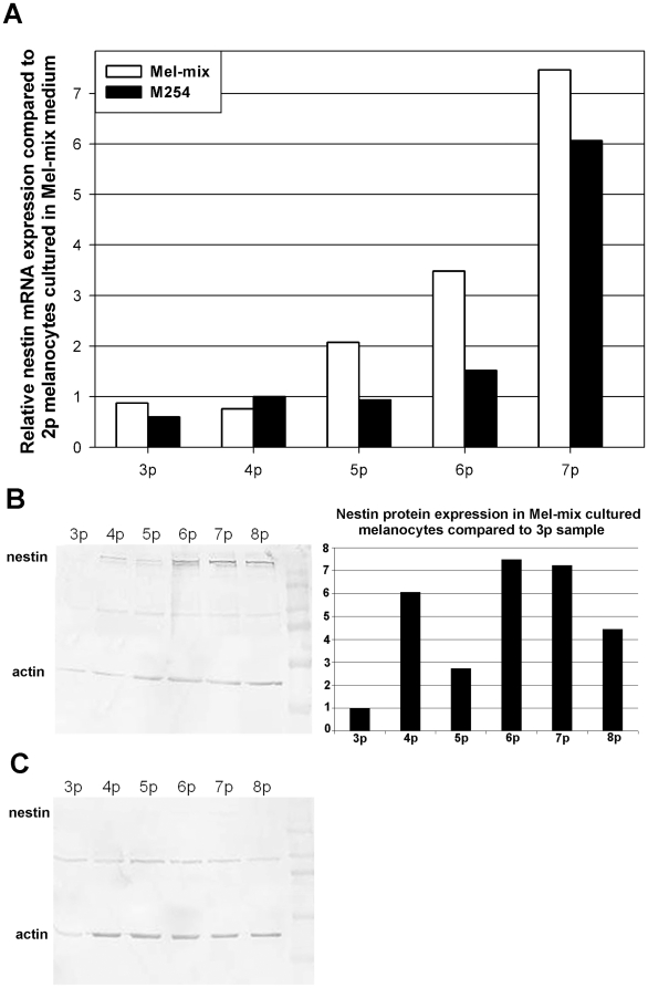
Figure 7. Nestin is strongly expressed in dedifferentiated melanocytes.
Although nestin mRNA increased with increasing passage number in all cultured cells this increase was more pronounced in dedifferentiated melanocytes (A). Using a monoclonal antibody, nestin was easily detactable in dedifferentiated cells (B), while in differentiated melanocytes only weak specific bands, not detectable by densitometry, were visible on the blot (C). Densitometry showed increasing expression in dedifferentiated cells during culturing (B).

