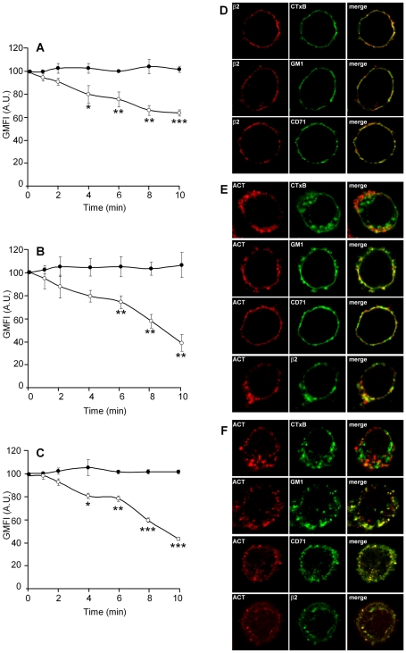
Figure 1. ACT is internalized and it triggers endocytosis of integrins and cholesterol-rich membrane microdomains in J774A.1 cells.
Addition of ACT (35 nM) to J774A.1 cells results in time-dependent internalisation of ACT (A), GM1 (B) and the β2 integrin (C) ([•] control cells and [○] ACT-treated cells). Analysis by confocal microscopy of the localization of ACT, GM1 and the β2 integrin in J774A.1 cells treated for 2 min at 37°C with 35 nM toxin (D) and cells treated for 10 min at 37°C with the same toxin concentration (E). Internalisation in A, B and C was analysed with FACS as described in Materials and Methods. The data shown are the mean ± SEM of at least three independent experiments, with *p<0.05, **p<0.025 and ***p<0.001.

