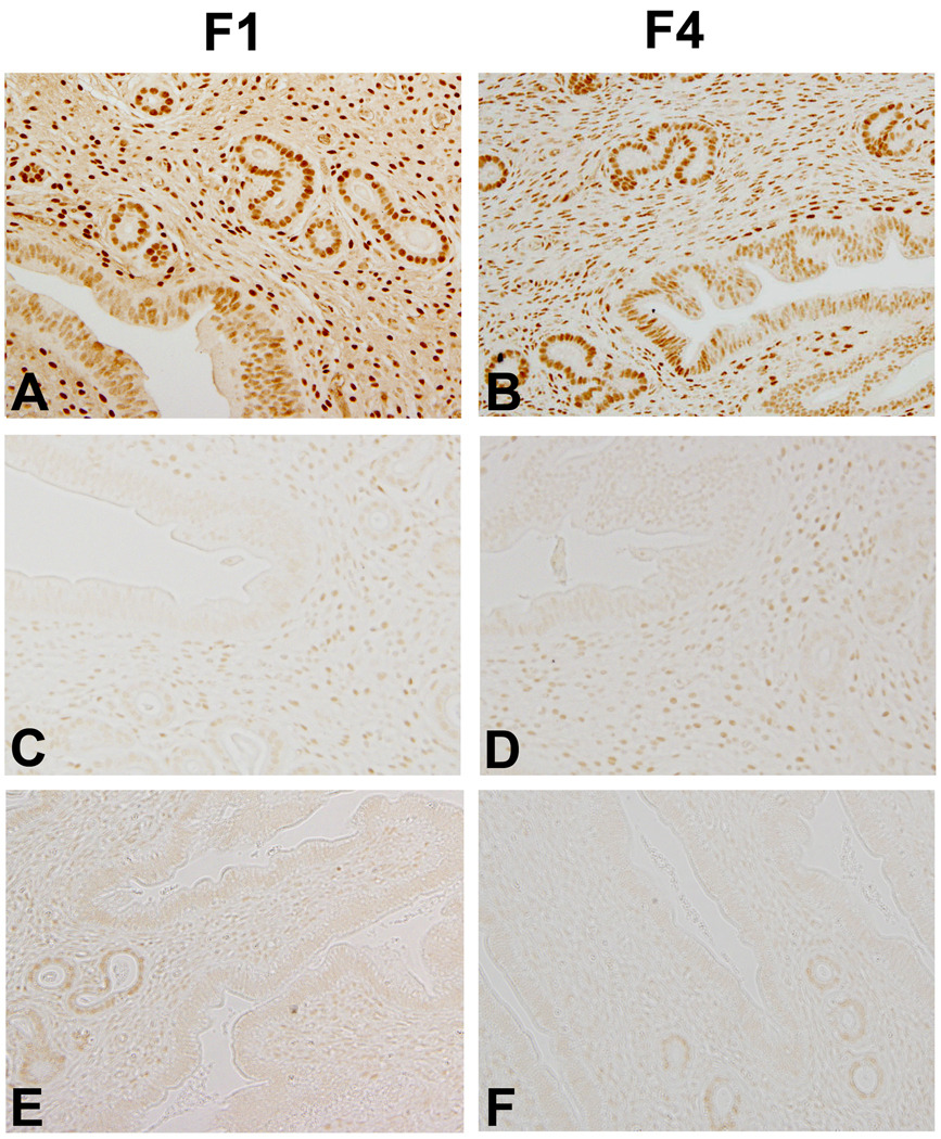
Figure 1. Immunolocalization of PR in Uteri of Adult Mice.
(A) Vehicle-exposed control F1 (conF1) mouse euthanized during estrous exhibits abundant PR immunolocalization; (B) PR immunolocalization of a conF4 mouse descended from a conF1 mouse also reveals abundant PR immunostaining. (C) PR immunolocalization in an infertile F1 female exposed to TCDD in utero only demonstrates reduced stromal and epithelial cell PR expression. (D) PR immunostaining of a uterus from an infertile F4 mouse, descended from a fertile F1 mouse which was exposed to TCDD in utero. (E) PR immunolocalization in the uteri of an infertile F1 female exposed to TCDD in utero and just prior to puberty reveals minimal PR expression in the stromal and epithelial compartments. (F) PR immunolocalization in the uterus infertile F4 female descended from a dually-exposed F1 female also exhibits a greatly reduced expression of PR protein in all compartments. Original magnification, 20×. Images are representative of results from at least four mice per group.

