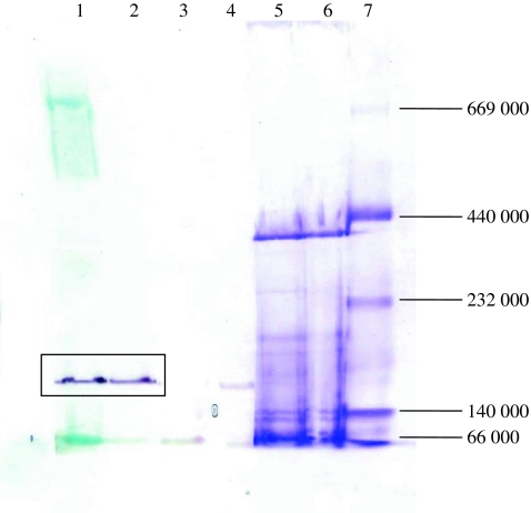
Fig. 2.
Activity staining of the native gel. Lanes 1–4, cell extract stained by different substrates, methylene blue and NBT (lane 1: 2-cyclohexenone, lane 2: 3-hydroxycyclohexanone, lane 3: cyclohexanol, lane 4: cyclohexanone); lanes 5 and 6, cell extract stained by Coomassie brilliant blue R-250; lane 7, protein marker (albumin (66,000), lactate dehydrogenase (140,000), catalase (232,000), ferritin (440,000), thyroglobulin (669,000))

