
FIGURE 2.
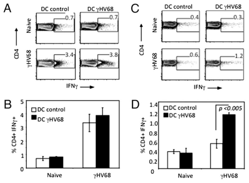
CD4 T cell production of IFN-γ is γHV68 specific. CD4 T cells were isolated from spleens of noninfected controls or long-term γHV68-infected mice and purified by negative selection. The purified CD4 cells were stimulated immediately with DCs that had been pulsed with control 3T3 lysates (DC control) or pulsed with lysates of 3T3 fibroblasts infected with γHV68 (DC γHV68) (A, B) or rested overnight in culture media and then stimulated as described above (C, D). Five hours after stimulation, intracellular cytokine staining of IFN-γ production by the CD4 splenocytes was measured by FACS. Error bars represent SD.