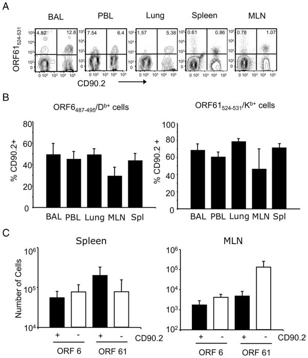
FIGURE 3.
γHV68-specific memory CD8 T cells mount a recall response to γHV68 infection. Sublethally irradiated B6.PL mice received 1 × 10⁶ CFSE-labeled CD44+CD8+ T cells isolated from the spleens of B6 mice that have been infected with γHV68 for at least 3 mo. The recipient B6.PL mice were rested for 30 days after adoptive cell transfer and subsequently infected with γHV68. On day 14 after infection, we analyzed bronchoalveolar lavage (BAL), peripheral blood lymphocytes (PBL), lung, MLN, and spleen of individual mice. A, Representative FACS plots showing tetramer-positive (ORF61524–531/Kb) cells of donor origin (CD90.2+) after gating on CD8+ T cells. Numbers indicate the frequency of tetramer-positive cells of donor (upper right quadrant) and host (upper left quadrant) origin. B, Bar diagrams show the frequency of cells of donor origin (CD90.2+) among virus-specific ORF6487–495/Db+ CD8+ T cells (left panel) or ORF61524–531/Kb+ CD8+ T cells (right panel). C, Bar diagrams represent the number of virus-specific CD8 T cells (ORF6487–495/Db+ and ORF61524–531/Kb+) that are of donor origin (CD90.2+) or host cells (CD90.2−). Left panel, Spleen; right panel, MLN. Bars represent the mean value of three experimental animals, and error bars indicate SEM. Similar results were obtained in two independent experiments.

