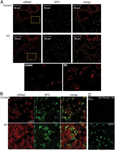
Fig. 3.
Increased ENaC protein expression in lung epithelia and type II cells from Nedd4L conditional KO mice. (A) Immunostaining of cryo sections from lungs of 10-day-old Nedd4L KO (Nedd4Lf/f;SPC-rtTA;teto-Cre) and control (Nedd4Lf/f;SPC-rtTA) mice stained with anti αENaC antibodies (red) that are directed to the extracellular domain of αENaC. Tissues were then permeabilized and stained with anti-SPC antibodies (green). (Bottom) Enlargements of the insets indicated, revealing increased ENaC immunoreactivity in alveolar-type II cells of the KO lungs. (B) Immunostaining of isolated, nonpermeabilized type II cells with anti-αENaC antibodies followed by permeabilization and staining for SPC and analysis by confocal microscopy, as in A. Note the increase in membrane staining of ENaC in the KO type II cells relative to controls. (C) Immunostaining controls showing lack of staining of type II cells in the absence of primary anti-αENaC antibodies (Upper). (Lower) Depicts SPC staining of the same cells (after permeabilization). Immunostaining of lung sections and isolated type II cells was carried out using lungs from 4 KO and 7 control mice, with representative micrographs shown.

