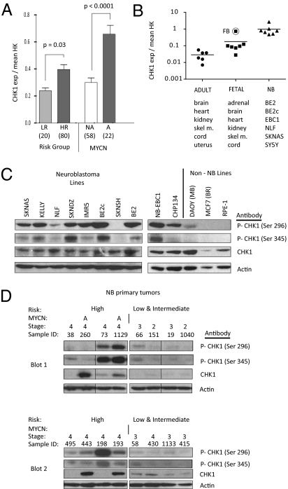
Fig. 3.
CHK1 mRNA and protein are highly expressed in neuroblastoma. (A) mRNA expression profiling of 100 neuroblastoma diagnostic tumors showed that CHK1 is overexpressed in HR tumors compared with LR tumors (Left) and in MYCN-amplified tumors compared with MYCN NA tumors (Right) (mean ± SEM). HR, high-risk; LR, low-risk; NA, single-copy. (B) CHK1 mRNA expression by real-time PCR demonstrates that CHK1 is overexpressed in neuroblastoma cell lines and embryonal tissues compared with adult tissues. (C) Western blot with constitutive phosphorylation of CHK1 Ser296 and Ser345 in neuroblastoma cell lines compared with control cell lines. (D) Western blot of phosphorylation of CHK1 Ser296 and Ser345 and CHK1 in neuroblastoma primary tumors. A, genomic amplification of MYCN (samples 260 and 1,129). Each of the two blots (blot 1 and blot 2) had four low/intermediate-risk and four high-risk tumors run in parallel. Fig. S6 shows the original order of blot 1, and blot 2 is ordered as in the figure.

