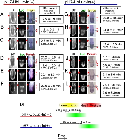
Fig. 2.
Introns delay the timing of Hes7 reporter expression. (A–L) Comparison of reporter expression (Luc) with either Hes7 intron expression or Hes7 protein expression in pH7-UbLuc-In(−) mice (A–F) or pH7-UbLuc-In(+) mice (G–L). Reporter expression was classified into three phases on the basis of luciferase activity: phase A, relatively narrower expression in the anterior PSM and broader expression in the posterior PSM (A, D, G, and J); phase B, strong expression in the middle part of the PSM (B, E, H, and K); and phase C, relatively broader expression in the anterior PSM and narrower expression in the posterior PSM (C, F, I, and L). After acquisition of luciferase activity images, posterior parts of the embryos were fixed immediately and analyzed for either Hes7 intron expression (n = 16 in A–C, n = 7 in G–I) or Hes7 protein expression (n = 22 in D–F, n = 22 in J–L). The first column shows bright field (BF) images. The difference in space (1 unit is defined as a one-somite length) was converted into the difference in time on the basis of the movies. The averages with SEs are shown. The arrowhead on the left side of each section indicates a boundary between a newly formed somite and the PSM. (M) Comparison of Hes7 reporter expression with Hes7 gene transcription and Hes7 protein expression.

