Fig. 5.
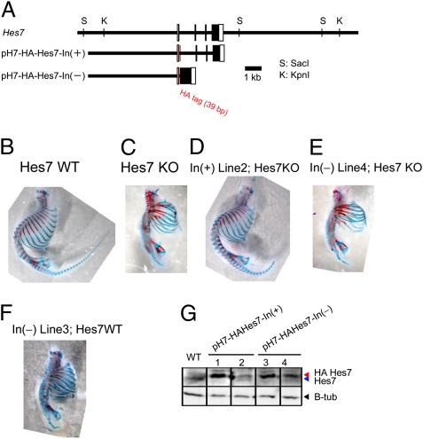
Segmentation defects of Hes7-null mice were rescued by the intron(+) Hes7 transgene but not by the intron(−) Hes7 transgene. (A) Structures of intron(+) and intron(−) Hes7 transgenes. The HA tag was added to the amino terminus to differentiate between the endogenous and transgene expression. This tag did not affect the segmentation (D). (B) Wild-type mouse. (C) Hes7-null mouse. (D) Hes7-null mouse containing the intron(+) Hes7 transgene. Four independent lines containing the intron(+) Hes7 transgene including the one shown here (line 2) rescued the segmentation defects. (E) Hes7-null mouse containing the intron(−) Hes7 transgene. Four independent lines containing the intron(−) Hes7 transgene including the one shown here (line 4) did not rescue the segmentation defects. (F) Segmentation defects were caused even in the Hes7(+/+) backgroud by the intron(−) Hes7 transgene (line 3). Bones and cartilages of neonates were stained in red and blue, respectively (B–F). (G) Western blot analysis of the PSM in the Hes7(+/+) background. Hes7 protein levels in two independent lines of the intron(+) transgene (lines 1 and 2) and two independent lines of the intron(−) transgene (lines 3 and 4) are shown. Hes7 protein made from the transgene is larger in size than the endogenous one because of the HA tag. β-Tubulin was used as a control. Note that the endogenous Hes7 protein expression was down-regulated by the exogenous Hes7 protein expressed from the transgenes. Segmentation defects of Hes7-null mice were rescued by the intron(+) transgene lines 1 and 2 but not by the intron(−) transgene lines 3 and 4.

