Fig. 1.
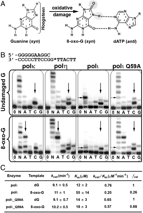
Activity of 8-oxo-guanine and human Y-family DNA polymerases. (A) Structural changes of guanine to 8-oxo-G. Bracket emphasizes the Hoogsteen edge and gray circles represent sites of modification on 8-oxo-G. The mismatched OG∶dATP base pair is shown on the right. (B) Primer extension assays show differences in nucleotide incorporation by polκ, polη, polι, and polιQ59A mutant for undamaged G (Upper) and 8-oxo-G (Lower) bases. The DNA substrate used for replication assays is shown at the top with G* representing the site of modification. Vertical black arrows indicate nucleotide insertion preference, whereas horizontal black arrows indicate replication stalling. Enzymes were incubated with DNA in the absence of nucleotides (0), presence of all four nucleotides (N), or individual nucleotides (A, T, C, G). (C) Steady-state kinetic parameters for dCTP incorporation opposite undamaged G (dG) and 8-oxo-G by wild-type polι and the polιQ59A mutant. The relative incorporation efficiency between damaged and undamaged DNA is reported as frel = (kcat/Km)damaged/(kcat/Km)undamaged.

