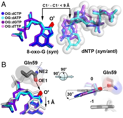
Fig. 2.
Positioning of 8-oxo-G∶dNTP in the Polι/DNA/nucleotide ternary structures. (A) Superposition of replicating base pairs from all four polι/8-oxo-G/nucleotide ternary structures: OG∶dCTP (blue), OG∶dATP (cyan), OG∶dGTP (purple), and OG∶dTTP (pink). The oxygen modification on 8-oxo-G is colored red and the C1′–C1′ distances are indicated with black arrows. Sphere representation is shown for the incoming nucleotides. (B) Positioning (Left) and tilting (Right) of the 8-oxo-G bases by Gln59. The 8-oxo-G bases are superimposed with a previous template G polι structure (PDB ID 2ALZ, gray) with a black arrow indicating repulsion between the OE1 atom of Gln59 and the O8 atom of 8-oxo-G and a dashed line on the NE2 atom representing a hydrogen bond with a backbone carbonyl oxygen. The template bases are numbered 0 and the underlying bases are numbered -1. Gray dashed lines on the right indicate the planes of the guanine bases.

