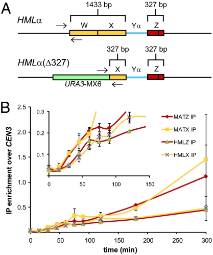
Fig. 4.
Both sides of a DSB at MAT interact with homologous HML donor sequences. (A) Schematic of WT HML and HML(Δ327) showing the extent of homology shared with both sides of an HO-induced DSB at MAT. Arrows indicate PCR primer positions used to detect Rad51-mediated synapsis between the left side of a DSB at MAT and HML. (B) Kinetics of Rad51 ChIP to the indicated regions of MAT and HML during MAT switching in HML(Δ327) cells. Inset: Graph is enlarged part the larger graph to show differences in the timing of Rad51 recruitment to MAT and HML(Δ327). Rad51ChIP data are normalized to CEN3. Data represent three independent experiments; error bars indicate SEM.

