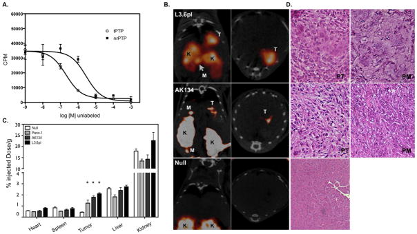
Figure 3. In vivo imaging of Plec1 in orthotopic PDAC.
A) In vitro validation of tPTP. L3.6pl cells were plated on a 96-well plate and incubated with 111In-tPTP and increasing log concentrations of unlabeled tPTP or negative control tetramer. B) Mice bearing tumors from orthotopically implanted L3.6pl, AK134 cells and mice without tumors (null) were injected with 111In-tPTP and imaged via SPECT/CT 4 hours post injection. Note the accumulation of tPTP in PDAC, allowing the in vivo imaging of tumor in the pancreas and in peritoneal metastases. Coronal (left) and axial (right) SPECT/CT slices through the tumor are presented. T-tumor, K-kidney, M-peritoneal metastasis. C) After SPECT/CT imaging, animals were sacrificed, organs harvested and gamma counts assessed. Null data is from both nu/nu and FVB/NJ animals that were injected in the pancreas with saline. D) Histology. Animals that had orthotopically implanted tumors or null animals were sacrificed and pancreas and regions of visible peritoneal metastases were removed, embedded, sectioned and stained with H&E (20x image). PT–primary tumor, PM–peritoneal metastasis

