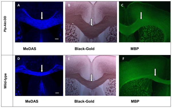
Figure 2.
In vitro MeDAS staining (A and D) of myelin sheaths in corpus callosum in comparison with Black-Gold (B and E) and MBP staining (C and F) in adjacent sections. Arrows show myelinated corpus callosum. A-C, Plp-Akt-DD mouse brain sections stained with MeDAS, Black-Gold and MBP, respectively. D-F, Control mouse brain sections stained with MeDAS, Black-Gold and MBP, respectively. The corpus callosum region visualized by MeDAS appeared to be much larger in the Plp-Akt-DD mouse brain than in the control littermate wild-type mouse brain, which was the same pattern that was observed in the Black-Gold or MBP antibody staining.

