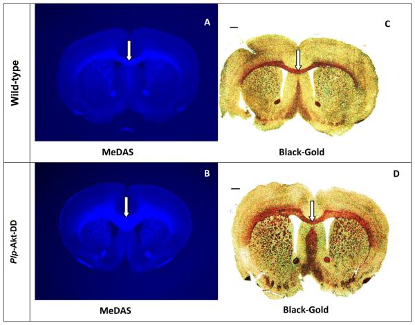
Figure 4.
In situ MeDAS staining of myelin sheaths in the corpus callosum in correlation with Black-Gold staining. (A, B) In situ staining of MeDAS in the control (A) vs hypermyelinated mouse brains (B). (C, D) Black-Gold staining in the control (C) and hypermyelinated mouse brains (D) in adjacent sections, respectively. In the corpus callosum of Plp-Akt-DD mice, MeDAS staining readily detects the enhancement of myelination, which had resulted in the enlargement of the corpus callosum region. Arrows show myelinated corpus callosum.

