Figure 1.
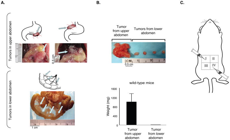
Inoculation of CT26 tumor cells into the peritoneal cavity leads to differential tumor growth in the upper versus lower abdomen. A. Upper panel: CT26 carcinoma cells form large, angiogenic tumors in the upper region of the peritoneal cavity and small, non-angiogenic tumors on the mesentery in the lower abdomen. Arrows point to large angiogenic tumors located on or below the stomach in upper panel, and to small non-angiogenic tumors in the lower abdomen (n=10 mice per group). B. The size and weight of CT26 tumors isolated from the upper abdomen are significantly larger than tumors from the lower abdomen. Data are represented as mean +/− SEM. C. Schematic depicting the sites of tumor cell inoculation (quadrants I and IV) and the direction of the needle.

