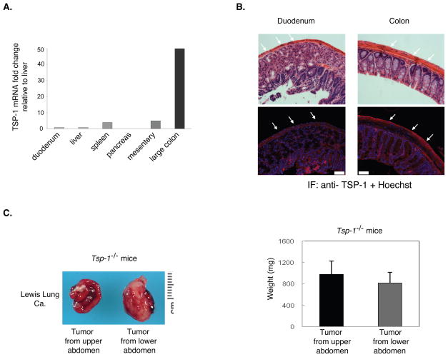
Figure 3.
Differential expression of thrombopsondin-1 (TSP-1) in the stroma of the upper and lower abdomen. A. Tsp-1 mRNA expression in organs from the upper and the lower abdomen. The fold change is relative to liver and normalized to GAPDH. B. TSP-1 immunofluorescence of the serosal surface of organs in the upper (duodenum) versus lower (colon) abdomen. White arrows identify the serosal surfaces. Upper panel: H&E. Lower panel: Anti-TSP-1 (red) immunofluorescence and Hoechst dye (blue). Pictures are taken at 63×. Scale bar = 50 μm. C. The differential growth of Lewis lung tumors in the upper versus lower abdomen is lost in Tsp-1−/− mice. The weight of tumors isolated from the upper (small intestine) and lower (large colon) abdomen was measured (n= 10 mice per group). Data are represented as mean +/− SEM.

