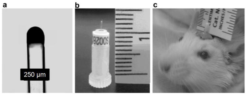
Fig. 1.

The implantable optode (a) micrograph of the fiber optic probe (10X). (b) Photograph of a chronically implantable probe supported by a transport sleeve. (c) Photograph of implanted fiber optic probe showing size, location and the supporting ring of dental cement. The picture was taken when the animal was awake.
