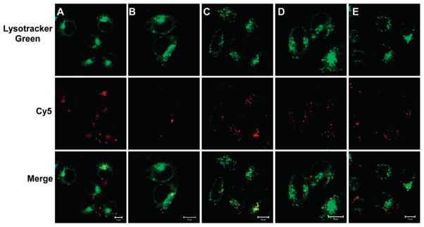
Figure 7.
CLSM images of HeLa cells transfected by PEI/PAH-Cit/PEI/MUA-AuNPs (A), Lipofectamine (B), PEI (C), PEI/MUA-AuNPs (D), and PEI/PSS/PEI/MUA-AuNPs (E). The w/w ratio of Au/DNA 10.0 was used for all of the gold nanoparticles. Images were taken after cells were incubated with NPs for 6 h. siRNA was labeled with cy5 (red, middle row). The late endosome and lysosome were stained with LysoTracker Green (green, first row). The yellow fluorescence (third row) is a result of colocalization of LysoTracker Green and cy5-labeled siRNA. The scale bar is 10 μm.

