
Figure 4. Ndel1 Function is Essential for Migration of Olfactory Interneurons.
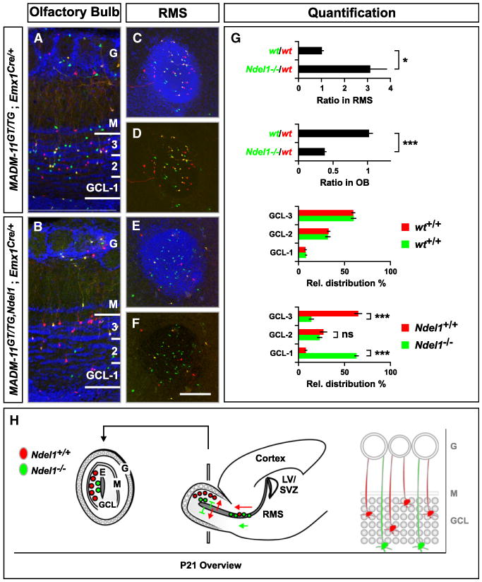
(A and B) Distribution of olfactory bulb interneurons (oINs) across the granule cell layer (GCL) in the P21 OB. Genotypes are indicated, Ndel1−/− cells labeled with GFP (green) and WT cells with tdT (red). Note the reduction of green Ndel1−/− cells in (B).
(C–F) Distribution of migrating oINs in cross sections of the RMS. Nuclei were stained using DAPI to outline the cytoarchitecture of the OB (A and B) and RMS (C and E). G, glomerular layer; M, mitral cell layer. Scale bar, 100 μm (A and B); 150 μm (C–F).
(G) Quantification of the green/red ratio in the RMS and the OB (upper panels) and the relative distribution (%) of oINs across three equal sectors in the GCL (lower panels). Values represent mean ± SEM ns: nonsignificant; *p < 0.05 and ***p < 0.001. (H) Schematic summary of migration of WT (red) and Ndel1−/− (green) MADM-labeled oINs. Red WT oINs originate from the subventricular zone of the lateral ventricle (SVZ/LV), migrate along the RMS to the OB, where oINs exit the RMS to migrate centrifugally to occupy different layers of the GCL. Green Ndel1−/− oINs accumulate along the RMS as indicated by the higher number of green circles in the RMS, and accumulate significantly closer to the ependymal layer (E) (extension of RMS) within the OB than red WT cells, suggesting a defect in migration in the target lamina (gray circles in the high magnification scheme on the right). G, glomerular layer; M, mitral cell layer; GCL, granule cell layer.
See also Figure S6.