
Figure 5. Ndel1 Is Required for Migration of Cerebellum Purkinje and Granule Cells.
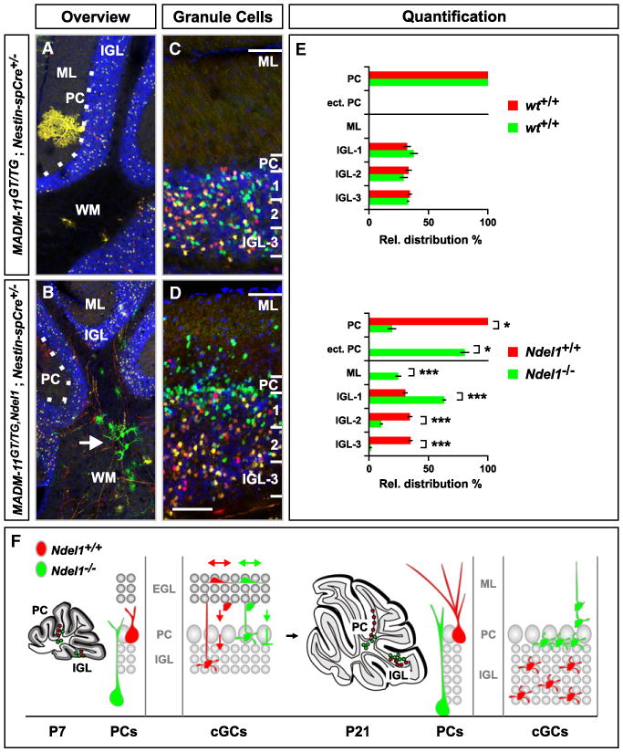
(A–D) Distribution of Purkinje cells and cGCs in P21 cerebellum. Genotypes are indicated, Ndel1−/− cells labeled with GFP (green) and WT cells with tdT (red). (A and B) Central part of the cerebellum with the white matter (WM), Purkinje cell (PC) layer (white dotted line), internal granule cell layer (IGL) and molecular layer (ML) labeled. Ndel1−/− Purkinje cells are mostly localized in the white matter (B, white arrow). See Figure 8F for high-resolution image of Ndel1−/− Purkinje cell. (C and D) Distribution of cGCs in control- (C) and Ndel1-MADM (D). Scale bar, 150 μm (A and B); 60 μm (C and D).
(E) Quantification of Purkinje cell distribution (%) in the PC layer or in ectopic locations (ect. PC), and cGCs across the molecular (ML) and internal granule layer (IGL), in control- (upper panel) and Ndel1-MADM (lower panel). The IGL was divided into three equal sectors for quantification of the relative distribution of cGCs. Values represent mean ± SEM ns: nonsignificant; *p < 0.05 and ***p < 0.001.
(F) Schematic summary of migration of WT (red) and Ndel1−/− (green) cerebellar Purkinje cells and cGCs. cGCs are born at the most superficial sublayer of the external granule layer (EGL) during the first 3 postnatal weeks (left panel). Nascent WT cGCs migrate inward across the ML, pass the PC layer, and settle throughout the IGL (small gray circles representing the final positions of cGCs). Most Ndel1−/− cGCs also migrate across the ML, but a fraction fails to pass the PC layer. Ndel1−/− cGCs that pass the PC layer accumulate at the most superficial sublayer of the IGL.
See also Figure S6.