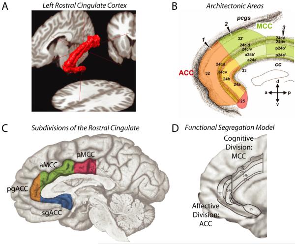
Figure 1. Divisions of the human rostral cingulate.
The rostral cingulate has been partitioned on physiological and anatomical grounds at spatial scales ranging from the macroscopic to the molecular. A. Three-dimensional rendering of the left rostral cingulate cortex. The cingulate, shown in red, was manually traced on a single subject’s magnetic resonance image (MRI). Much of the constituent cortical gray matter lies buried within the cingulate sulci, a fact not apparent from inspection of the mesial surface (for additional information, see Box 1 and the Supplement). B. Architectonic areas of the cingulate. Areas were defined209 on the basis of differences in microanatomy and neurotransmitter chemistry and, hence, differ somewhat from the classical descriptions of Brodmann and other pioneering neuroanatomists1, 210. Architectonic features provide one means of defining homologies across species183, 211. Adapted with permission from Ref. 209. C. The four major subdivisions of the rostral cingulate. Subdivisions were defined by Vogt and colleagues211 on the basis of regional differences in microanatomy, connectivity, and physiology. The supracallosal portion of the cingulate is designated the midcingulate cortex (MCC) and is divided into anterior (aMCC: green) and posterior (pMCC: magenta) subdivisions. The portion of the cingulate lying anterior and ventral to the corpus callosum is designated the anterior cingulate cortex (ACC) and is divided into pregenual (pgACC: orange) and subgenual (sgACC: cyan) subdivisions by the coronal plane at the anterior tip of the genu. Adapted with permission from Ref. 212 (for additional information, see the Supplement). D. The functional segregation model of Bush, Luu and Posner. On physiological and anatomical grounds, Bush et al.14 argued that the rostral cingulate consists of two functionally segregated regions: a rostroventral ‘affective’ division (ACC; originally termed ‘ventral ACC’) and a dorsal ‘cognitive’ division (MCC; originally termed ‘dorsal ACC’). Adapted with permission from Ref. 14.

