Figure 1.

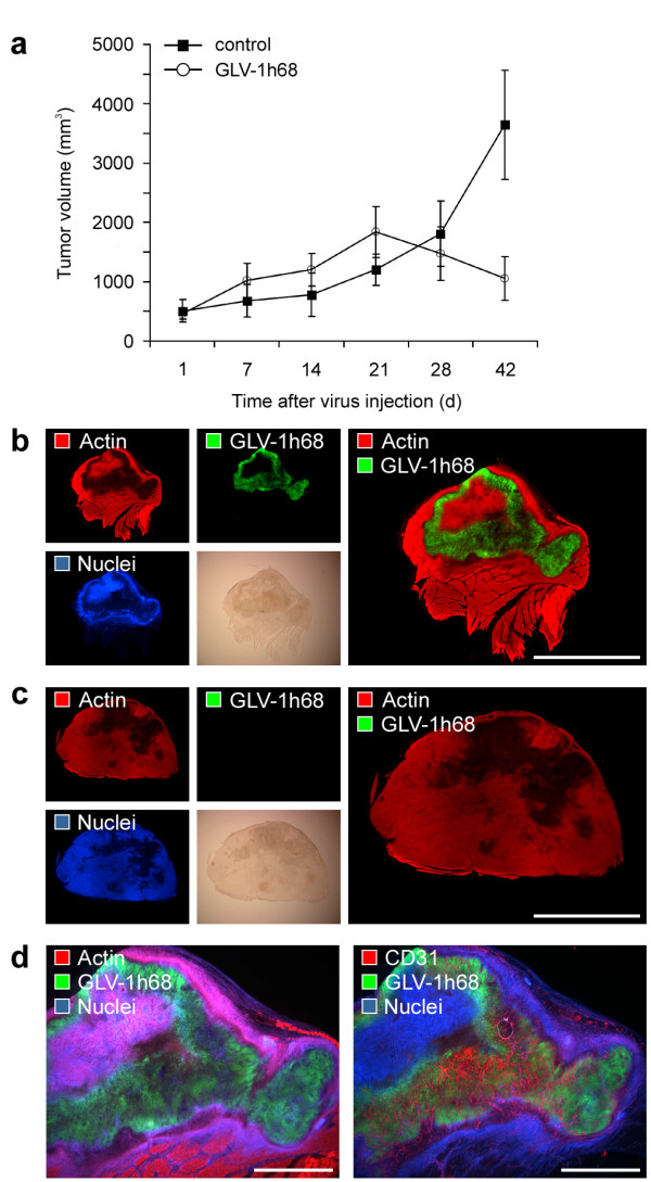
Oncolytic destruction of human GI-101A breast tumor xenografts. GI-101A tumor-bearing mice were intraveneously (i.v.) injected with 5 × 10⁶ pfu/100 μl PBS GLV-1h68 or PBS as a control. (a) Tumor growth was monitored weekly by measuring the tumor volume of six mice in each group. Shown are the mean values +/- standard deviations. The study was repeated in three independent experiments. (b, c) Whole tumor cross-sections (100 μm) of GLV-1h68-infected GI-101A (b) and control tumors (c) 42 days p.i. were stained with Phalloidin-TRITC (red) to label the actin cytoskeleton and Hoechst 33342 (blue) to visualize cellular nuclei; GFP fluorescence (green) indicated viral-infected cells. (d) Serial sections of whole tumor cross-sections of 42-days-colonized GI-101A tumors were labelled either with Phalloidin-TRITC (actin) or anti-CD31 antibody (red). In both sections nuclei were stained with Hoechst (blue) and GLV-1h68-infected cells were indicated by GFP fluorescence (green). The colonized, necrotic tumor tissue showed a dense CD31-positive vascular network. All images are representative examples. Scale bars represent 5 mm (b, c), 2 mm (d).
