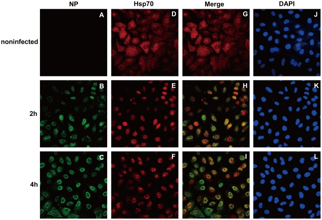
Figure 3. Hsp70 is translocated into the nucleus in infected A549 cells.
A549 cells were infected with rWSN at MOI of 5 for 1 h and cultured for indicated time. The infected and noninfected A549 cells were fixed and stained with rat anti-NP antibody (A, B and C) and rabbit anti-Hsp70 antibody (D, E and F), colocalization of Hsp70 and NP were shown in merged images (G, H and I), and DAPI staining indicated the nucleus (J, K and L).

