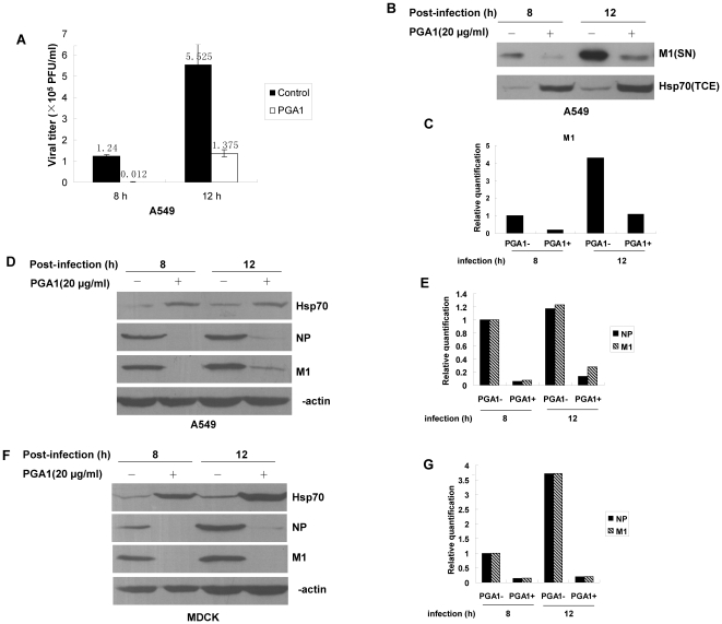
Figure 4. PGA1 treatment inhibits the propagation of influenza A virus.
Cells were infected with rWSN at MOI of 1, then cultured in the absence or presence of PGA1 for the indicated intervals. The supernatants and total cell extracts were harvested. (A) The supernatants from infected A549 cells were subjected to plaque assay to measure the viral titers shown as the data on the bars. Error bars represent standard deviation (n = 3). (B, C) M1 protein (SN) and Hsp70 (TCE) were detected by immunoblotting (B) with mouse anti-M1 and rabbit anti-Hsp70 antibodies (SN and TCE stand for supernatant and total cell extract, respectively). (C) The relative quantification of M1 in (B). (D–G) Total cell extracts of infected A549 cells (D, E) and MDCK cells (F, G) were prepared for immunoblotting with rabbit anti-Hsp70, rat anti-NP, mouse anti-M1, and rabbit anti-β-actin antibodies (D, F). (E, G) The relative quantification of M1 and NP in (D, F).

