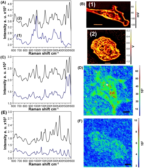
Figure 5. SERS monitoring at different cell cycle stages.
(A) SERS spectra of (black) resting HMEC cells (G0/G1) and (blue) mitotic HMEC cells (S/G2). (B) Confocal images of HMEC cells in the (1) resting state (G0/G1) and (2) mitotic state (S/G2), (C) SERS spectra of HMEC cells from cytoplasm (blue curve) and the nucleus (black curve), and (D) SERS map image of HMEC cells. (E) SERS spectra of HepG2 cells from cytoplasm (blue curve) and the nucleus (black curve), and (F) SERS map image of HepG2 cells. Scale bar: 10 µm.

