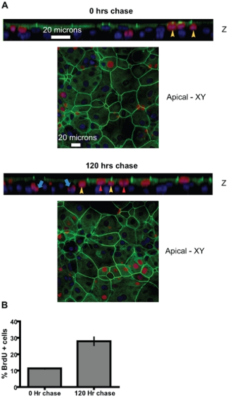
Figure 2. Suprabasal multipotent cells give rise to both luminal and basal cells.
A BrdU pulse-chase experiment in MCF10A Transwell® cultures show the location of dividing epithelial cells. The MCF10A transwell® cultures were labeled with BrdU for 24 hours followed by medium change and continued cultivation of the cultures for the designated periods of time. (A) Representative confocal Z-section and apical XY-sections immunofluorescently stained for BrdU (red), ZO1 (green) and nuclei (blue). Note the suprabasal location of BrdU + cells (yellow arrowheads) at 0 h of chase. After 120 hours chase the BrdU label is seen in both the luminal cells (ZO1+) (red arrowheads) and the basal cells (blue arrows). Suprabasal putative multipotent cells found wedged between the luminal and basal layers are also observed (yellow arrowheads). (B) Quantification of BrdU + cells after 0 hours chase and 120 hours chase indicates a 2.5 fold increase in BrdU + cells. The data is expressed as means +/− S.E.M.

