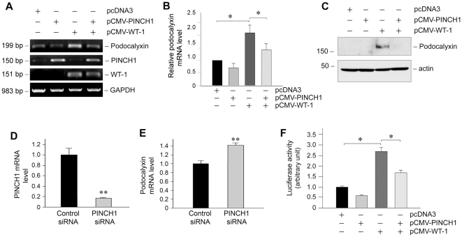
Figure 7. PINCH1 blocks WT1-mediated podocalyxin expression in human podocytes.
A, RT-PCR analyses demonstrate that PINCH1 blocked WT1-stimulated podocalyxin mRNA expression in podocytes. Cells were transfected with expression vectors for PINCH1, WT1 or both, respectively. RT-PCR amplification of housekeeping GAPDH was performed in an identical manner to serve as controls. B, Graphic presentation shows the relative PINCH1 mRNA abundance in different groups after normalization with GAPDH. Data are presented as mean ± SEM of three independent experiments. *P<0.05. C, Western blot analyses show that PINCH1 blocked WT1-mediated podocalyxin protein expression. Human podocytes were transfected with different plasmids as indicated for 48 h. Total cell lysates were immunoblotted with specific antibodies against podocalyxin and actin, respectively. D and E, Knockdown of PINCH1 in podocytes promotes podocalyxin expression. Human podocytes were transfected with either control or PINCH1-specific siRNA. The expression of PINCH1 (D) and podocalyxin (E) was assessed by quantitative RT-PCR. **P<0.01 (n = 3). F, PINCH1 represses WT1-activated podocalyxin gene promoter activity. Human podocytes were co-transfected with different plasmids as indicated with luciferase- podocalyxin gene promoter reporter construct (pGL3-podocalyxin) for 48 h. Equal amounts of DNA were present in each transfection. Data are presented as mean ± SEM of three independent experiments. *P<0.05.

