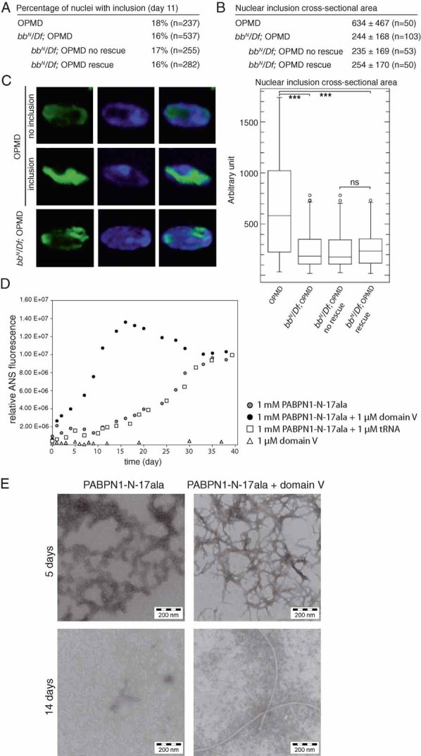
Percentages of nuclear inclusions in OPMD thoracic muscles at day 11, either in the presence or absence of bb deletions. In the presence of bb deletions, quantifications are indicated both regardless of the wing posture (bbN/Df; OPMD), and in subgroups showing or not wing posture defects (bbN/Df; OPMD no rescue, and bbN/Df; OPMD rescue)‘rescue’ indicates OPMD flies which have normal wing posture, ‘no rescue’ indicates flies which still have an affected wing posture.
Quantification of nuclear inclusion areas. Each nuclear inclusion was delimited in a focal plan and the surface area was calculated using Image J. Mean values of the surface areas are shown in arbitrary units (top). Distribution of nuclear inclusion surface areas are shown as box plots (bottom). Legend is as in
Fig 5B.
Confocal images of nuclear inclusions visualized with anti-PABPN1 staining. DNA was revealed with DAPI. Examples of nuclei without an inclusion (no inclusion), and with a large inclusion (inclusion) in OPMD thoracic muscles. Example of a small inclusion in an OPMD thoracic muscle in the presence of bb deletions.
Kinetics of fibril formation of the PABPN1-17ala N-terminal domain, either in the presence or absence of ribosomal RNA domain V, monitored by ANS fluorescence. tRNA was used as a negative control.
Electron micrographs of oligomers or fibrils of PABPN1-N-17ala formed in vitro, either in the presence or absence of ribosomal RNA domain V.