
Figure 2.
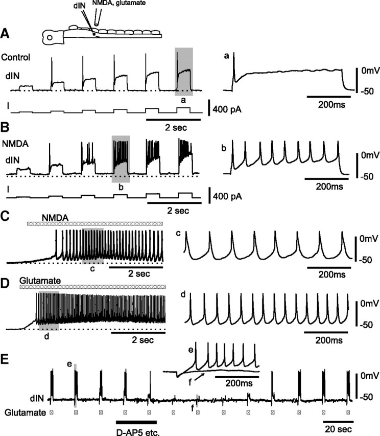
dINs fire repetitively to NMDA and glutamate application. A, At rest, a dIN fires single action potentials to current injections. B, dIN in A fires repetitively when 100 μm NMDA applied by microperfusion depolarized the membrane potential by ∼5 mV from rest (dotted line). C, Repetitive dIN firing response to microperfusion of NMDA alone. D, Another dIN fired repetitively to microperfusion of 1 mm glutamate. In C and D, saline contained 1 μm strychnine, 20 μm SR95531, 5 μm NBQX, 2 μm DHβe, and 150 μm Cd2+. E, The responses of a dIN to l-glutamate applied via a microiontophoresis electrode (−0.8 nA, ∼20 μm away from dIN) before, during, and after antagonist mixture (d-AP5, etc.: 50 μm d-AP5, 1 μm strychnine, 20 μm SR95531, 5 μm NBQX, 2 μm DHβe) dropped into a small well upstream to the recording chamber. Saline contained 150 μm Cd2+. Shaded area records a–e and f are expanded on the right.