
Figure 2.
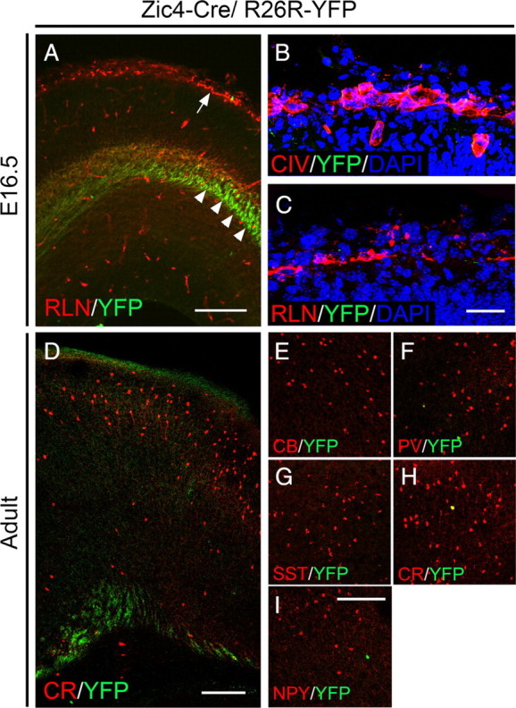
Absence of colocalization between YFP and molecular markers that label the meninges, C–R cells and interneurons in the neocortex in Zic4-CreTg/R26R-YFP+/− transgenic mice. A, E16.5 cortex showing expression of YFP and the C–R cell marker RLN. Some YFP-labeled thalamocortical fibers can be observed (arrowheads). The arrow points to C–R cells in the marginal zone where virtually no expression of YFP is observed. B, C, Absence of coexpression between YFP and Collagen IV (B) or RLN (C). D, Section through the adult cortex showing sparse, if any, YFP-expressing cells. Some YFP-labeled fibers can be observed. E–I, Immunolabeling for YFP and the interneuron markers CB, PV, SST, CR and NPY shows no or very little colocalization with all markers examined. Scale bars: A, 150 μm; B, C, 50 μm; D, 200 μm; E–I, 200 μm.