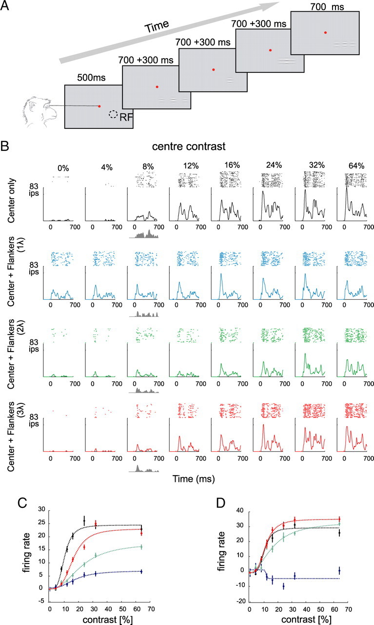
Figure 1.

Effects of collinear flankers on the activity of V1 neurons. A, The fixation task. The monkeys fixated a central fixation point (red) and passively viewed a sequence of four stimuli. Each stimulus consisted of a central target Gabor that was presented either in isolation or together with two collinear flankers. Stimulus presentation was 700 ms with an interstimulus interval of 300 ms. RF denotes receptive field location. B, Activity of a V1 neuron evoked by the presentation of the center stimulus alone or in the presence of collinear flankers located at one of three distances (1, 2, and 3λ). The gray shaded area represents the net response evoked by a target with a contrast of 8%. C, Contrast response function of the neuron shown in B. Note that the cell shows suppression at all contrast levels and every flanker distance, as was typical in our sample. The curves show fits of a hyperbolic ratio function. D, Contrast response function of a different, more exceptional neuron that showed some degree of facilitation for high-contrast stimuli in the presence of far (2 and 3λ) flankers. Error bars indicate SEM.
