Figure 8.
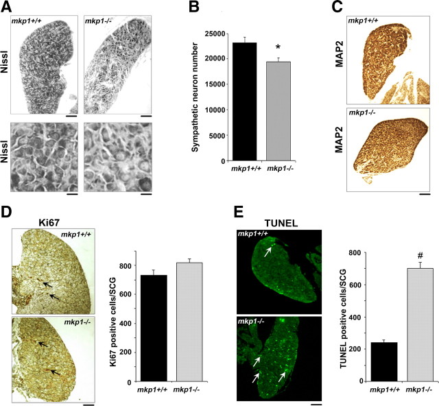
The number of sympathetic neurons is reduced during early postnatal development in mkp1−/− SCG compared to mkp1+/+ SCG. A, Brightfield images showing sections of mkp1+/+ and mkp1−/− P1 SCGs stained with Nissl. The top panel shows sections at low magnification (scale bar, 100 μm). The bottom panel shows a higher magnification (scale bar, 30 μm). B, Sympathetic neuron numbers in P1 SCGs isolated from mkp1+/+ and mkp1−/− mice were quantified in three animals of each genotype. Error bars represent SEM. *p < 0.02, Student's t test. C, Sections from mkp1+/+ and mkp1−/− P1 SCGs were also stained with an antibody to the neuronal marker MAP2. Scale bar, 100 μm. D, Sections from mkp1+/+ and mkp1−/− P1 SCGs were stained with the proliferation marker Ki67. Arrows indicate Ki67-positive cells. Scale bar, 100 μm. The number of Ki67-positive cells was determined in the mkp1+/+ and mkp1−/− P1 SCGs. Error bars represent SEM. p = 0.12. E, Sections from mkp1+/+ and mkp1−/− P1 SCGs were analyzed for apoptosis using TUNEL. Arrows indicate TUNEL-positive cells. Scale bar, 100 μm. The number of TUNEL-positive cells was determined in the mkp1+/+ and mkp1−/− P1 SCGs. Error bars represent SEM. p = 0.0004.

