Figure 1.

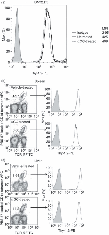
Thy-1 is expressed on a mouse invariant natural killer (iNKT) cell line and primary iNKT cells. DN32.D3 mouse iNKT cells were left untreated or stimulated with α-galactosylceramide (αGC; 100 ng/ml) for 24 hr. Cells were harvested and stained with phycoerythrin (PE)-conjugated anti-Thy-1.2 monoclonal antibodies (mAb) (open histograms) or isotype control (filled histogram). The expression of Thy-1 was analysed by flow cytometry and the mean fluorescence intensity was calculated (a). C57BL/6 mice were injected intraperitoneally with 2 μg αGC or vehicle. Splenocytes (b) and hepatic lymphoid mononuclear cells (c) were prepared 72 hr after injection. Cells were stained with a FITC-conjugated anti-T-cell receptor-β mAb, allophycocyanin-conjugated CD1d tetramer, and either PE-conjugated anti-Thy-1.2 mAb (open histogram) or isotype control (filled histogram). T-cell receptor-β+ CD1d tetramer+iNKT cells were gated on and Thy-1 expression was analysed.
