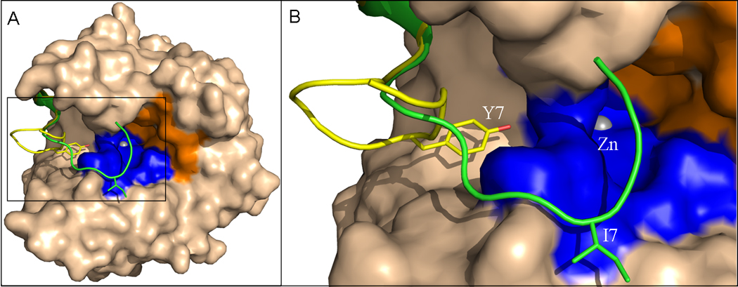
Figure 5.

Overall (A) and N-terminus (B) of superimposed crystal structures of wild-type HCA II and Y7I HCA II. The superimposed enzyme except the N-terminus is represented as a surface. The N-terminus of (yellow) wild type; and (green) Y7I HCA II is represented as ribbon, while the respective amino acids at position 7 as sticks. The hydrophobic and hydrophilic regions of the active-site are rendered orange and blue respectively. The active site zinc is depicted as a gray sphere.
