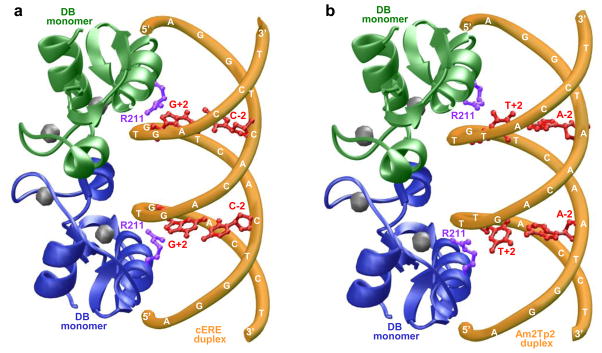
Figure 8.
3D atomic models of the DB domain of ERα in complex with dsDNA oligos containing the consensus ERE motif (a) and the variant Am2Tp2 motif (b). Note that the DB domain binds to DNA as a homodimer with a two-fold axis of symmetry. One monomer of the DB domain is shown in green and the other in blue. The Zn2+ divalent ions within each DB monomer are depicted as gray spheres and the sidechain moieties of R211 within each DB monomer are colored purple. The DNA backbone is shown in yellow and the bases at −2 and +2 positions within each motif are colored red.

