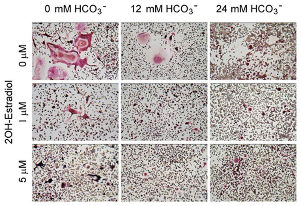
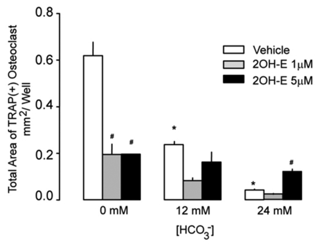
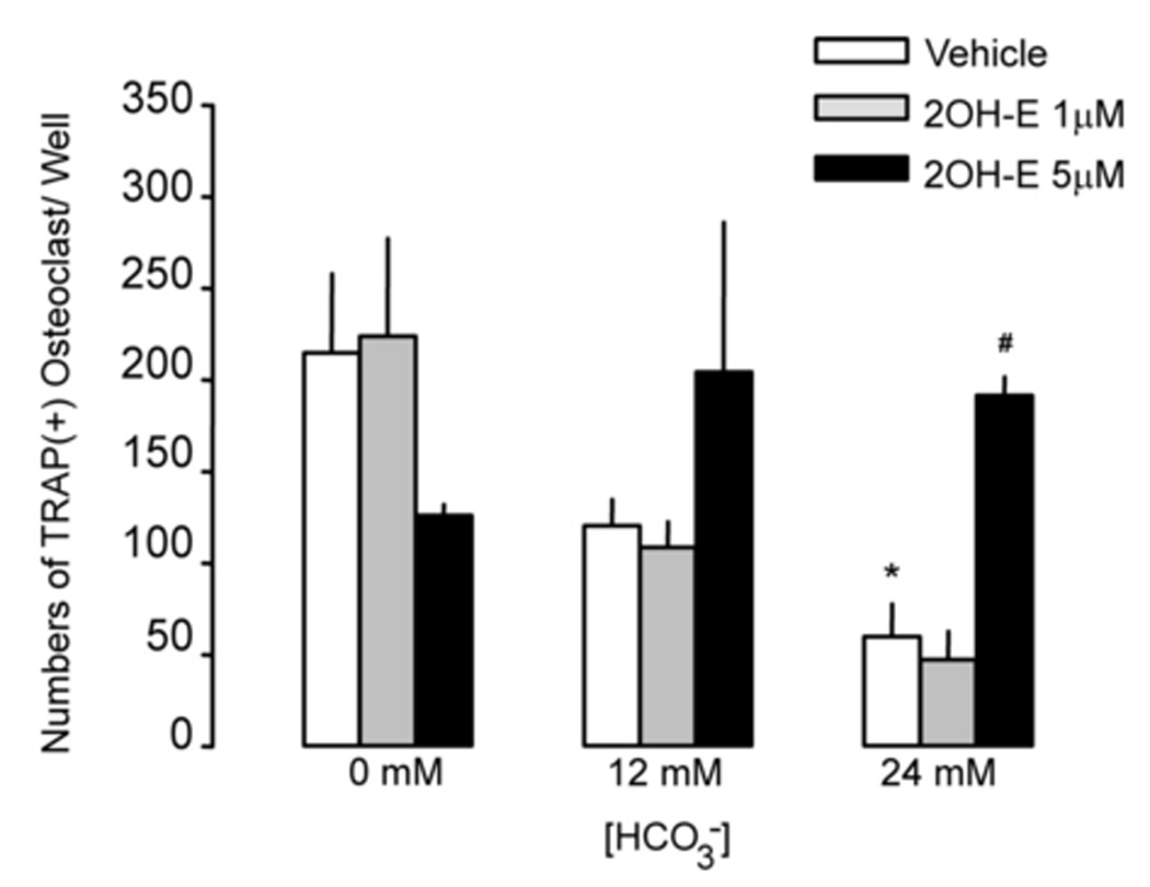
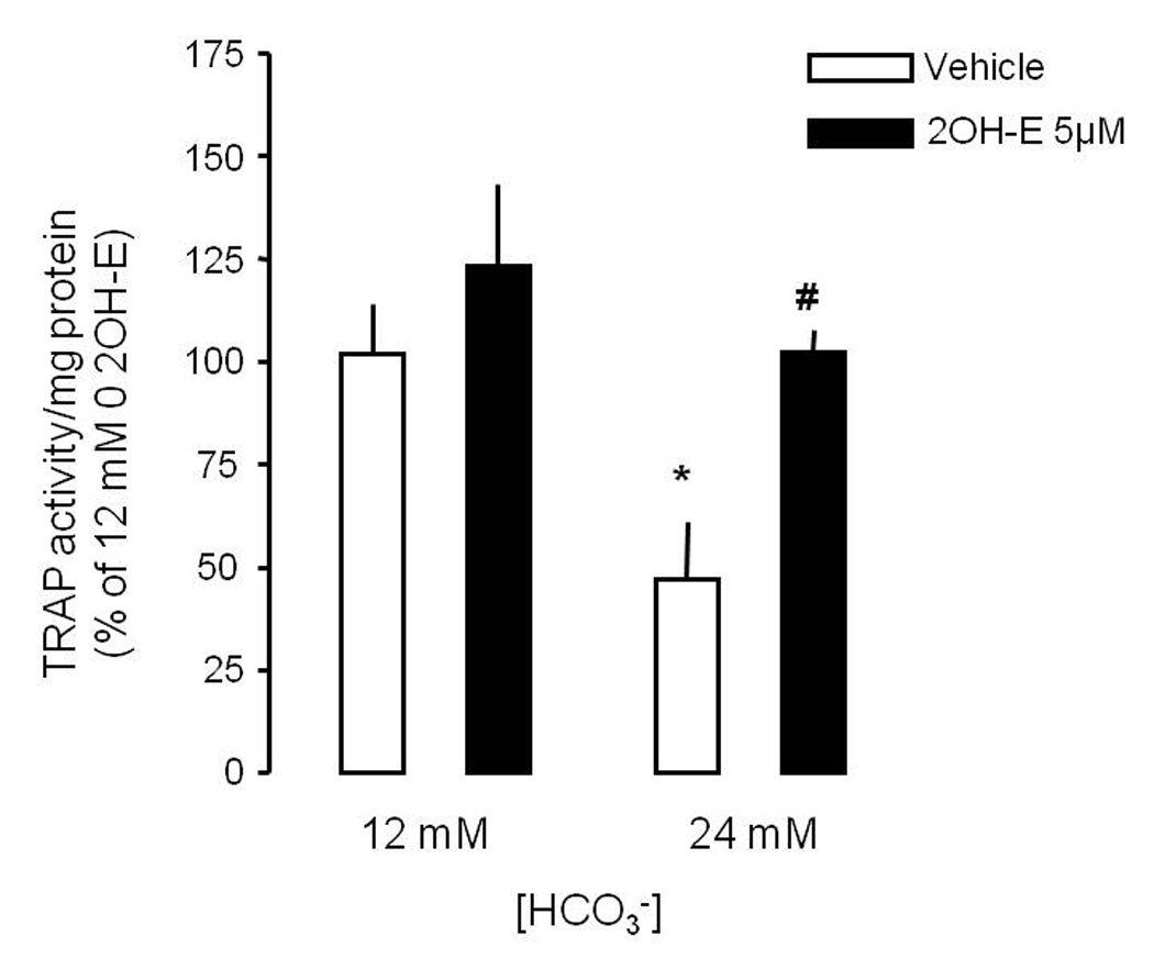
Fig. 1. Effects of HCO3− and sAC inhibitor on osteoclast formation.


A: RAW264.7 cells were seeded into the 24-well cell culture plates in DMEM media containing 50ng/ml RANKL and 5ng/ml M-CSF for induction of osteoclast differentiation. 0 mM NaHCO3, 12mM NaHCO3, 24 mM NaHCO3, +/− 1 µM, and 5 µM 2-hydroxyestradiol (all pH 7.4) were added for 7 days and cells were fixed and stained for TRAP (pink). Images show representative field from one of a total of four independent experiments. B: Effects of HCO3− on osteoclast size with 0 µM (open bar), 1 µM (gray bar), and 5 µM (black bar) 2-hydroxyestradiol. Four independent experiments were performed and in each one, four areas from each well were randomly selected for imaging. The area of TRAP (+)-multinucleated osteoclasts was determined using Image J software. Bars and error bars represent Means ± SEM from four separate experiments. C: Effects of HCO3− on osteoclast number with 0 µM (open bar), 1 µM (gray bar), and 5 µM (black bar) 2-hydroxyestradiol. Four independent experiments were performed and in each one, four areas from each well were randomly selected for imaging and TRAP (+)-multinucleated osteoclasts were counted and added together. * Significant difference compared to 0 mM HCO3− treated cells (p<0.05). # Significant difference compared to non 2-hydroxyestradiol treated cells (p<0.05)
