
Fig. 3. sAC and HCO3− regulate V-ATPase on osteoclasts.
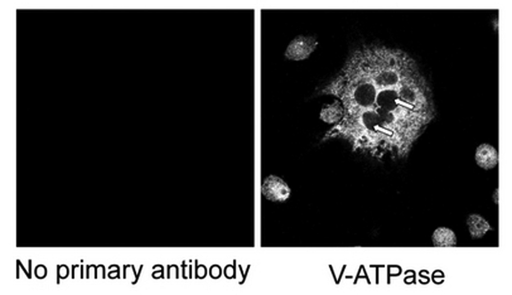
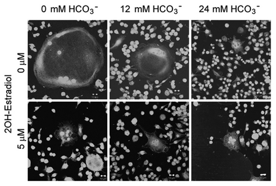
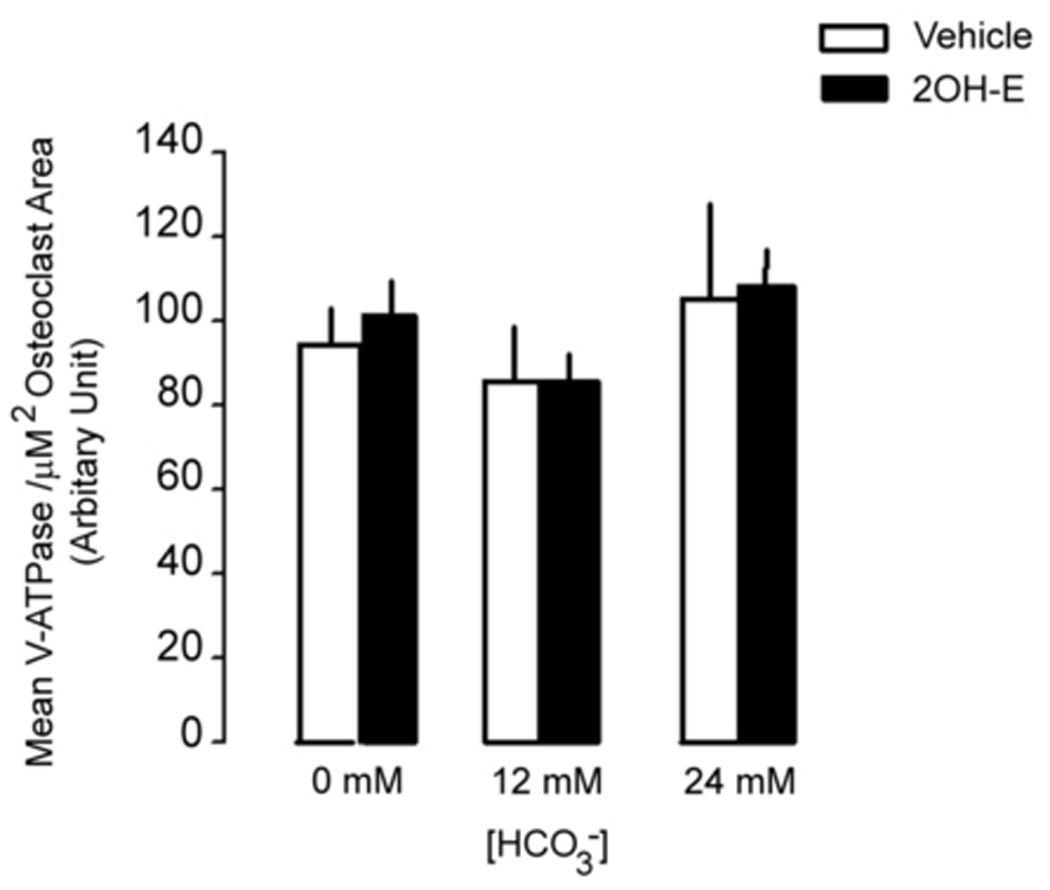
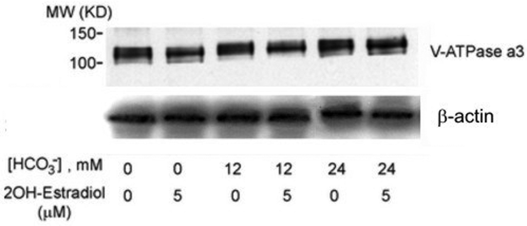
Osteoclasts were induced from RAW264.7 cells (50ng/ml RANKL and 5ng/ml M-CSF), fixed, permeablized, stained with anti-B subunit antibody, and visualized under confocal microscopy. A: V-ATPase is expressed throughout the cell in the cytosol but not in the nuclei (negative staining) of the multinucleated osteoclasts as well as the smaller RAW cells. No fluorescence signal was detected with secondary antibody alone (left panel). B: RAW264.7 cells were seeded into the cell culture plates with glass cover slips and induced with 50 ng/ml RANKL and 5 ng/ml M-CSF. 0 mM HCO3−, 12mM HCO3−, 24mM HCO3−, and +/− 5 µM 2-hydroxyestradiol were added into media for seven days. Cells were then fixed, permeabilized, stained with anti-B subunit antibody and visualized with confocal microscopy. Bar = 10 µm. C: Effects of HCO3− and 2-hydroxyestradiol on V-ATPase (B subunit) expression in single multinucleated osteoclast. Mean fluorescence intensity per µm2 area in osteoclasts was determined by Zeiss LSM image analyzer software. Bars and error bars represent means ± SEM. A total of 150 osteoclasts were randomly selected from 3 independent experiments. D: Effects of HCO3− and 2-hydroxyestradiol on osteoclast-specific a3 subunit expression in the total cell lysate of osteoclast. Induced osteoclasts were collected and 10 µg of total cell lysate was immunoblotted. β-actin served as loading control. One representative blot is shown; total of three showed similar results.