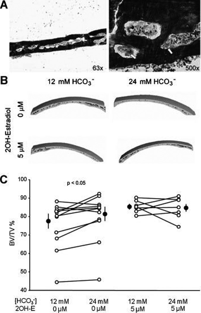
Fig. 6. Effect of HCO3− and sAC on bone mass in cultured mouse calvaria.

A: Toluidine blue staining of coronal section of mouse calvaria at low (left panel) and high (right panel) magnification from 8–9 week old mouse following one week culture. Osteoclasts (arrow) are attached to the trabecular bone surface. Image is one representative from eight calvaria analyzed. B: Three dimensional reconstructions of the partial mouse calvaria from µCT. Calvaria are cultured with 12mM or 24 mM HCO3− +/− 2-hydroxyestradiol for one week, then fixed with ethanol and µCT was performed. Images are representative of 16 mice. C: Line graphs summarize the HCO3− effect on the BV/TV% of mouse calvaria without (left panel) and with 2-hydroxyestradiol (right panel). Each line connects the two paired calvaria from the same mouse. Solid circles: mean ± SEM. P value is from paired t test.
